|
|
||
|---|---|---|
| .. | ||
| configs | ||
| datasets | ||
| tools | ||
| README.md | ||
README.md
Colorectal Nuclear Segmentation and Phenotypes (CoNSeP) Dataset
Description
This project supports Colorectal Nuclear Segmentation and Phenotypes (CoNSeP) Dataset, which can be downloaded from here.
Dataset Overview
The CoNSeP (Colon Segmentation and Phenotyping) dataset consists of 41 H&E stained image tiles, each with a size of 1,000×1,000 pixels and a magnification of 40x. These images were extracted from 16 colorectal adenocarcinoma (CRA) whole slide images (WSI), each of which belonged to a separate patient and was scanned using an Omnyx VL120 scanner at the Pathology Department of the University Hospitals Coventry and Warwickshire NHS Trust, UK. This dataset was first used in paper named, "HoVer-Net: Simultaneous Segmentation and Classification of Nuclei in Multi-Tissue Histology Images".
Original Statistic Information
| Dataset name | Anatomical region | Task type | Modality | Num. Classes | Train/Val/Test Images | Train/Val/Test Labeled | Release Date | License |
|---|---|---|---|---|---|---|---|---|
| CoNIC202 | abdomen | segmentation | histopathology | 7 | 4981/-/- | yes/-/- | 2022 | - |
| Class Name | Num. Train | Pct. Train | Num. Val | Pct. Val | Num. Test | Pct. Test |
|---|---|---|---|---|---|---|
| background | 27 | 83.61 | 14 | 80.4 | - | - |
| other | 17 | 0.17 | 9 | 0.52 | - | - |
| inflammatory | 25 | 2.66 | 14 | 2.14 | - | - |
| healthy epithelial | 3 | 1.47 | 2 | 1.58 | - | - |
| dysplastic/malignant epithelial | 10 | 7.17 | 8 | 9.16 | - | - |
| fibroblast | 23 | 3.84 | 14 | 4.63 | - | - |
| muscle | 8 | 1.05 | 3 | 1.42 | - | - |
| endothelial | 7 | 0.02 | 4 | 0.15 | - | - |
Note:
Pctmeans percentage of pixels in this category in all pixels.
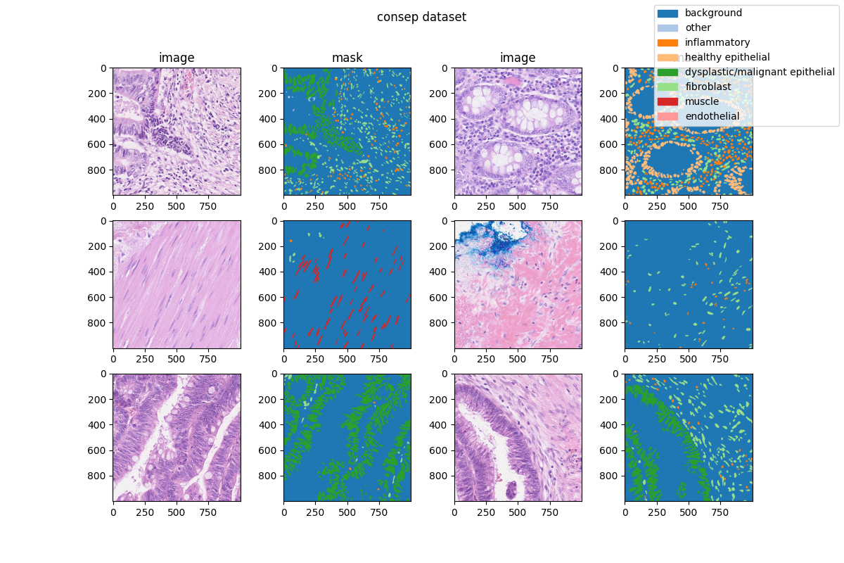
Visualization
Prerequisites
- Python v3.8
- PyTorch v1.10.0
- MIM v0.3.4
- MMCV v2.0.0rc4
- MMEngine v0.2.0 or higher
- MMSegmentation v1.0.0rc5
All the commands below rely on the correct configuration of PYTHONPATH, which should point to the project's directory so that Python can locate the module files. In conic2022_seg/ root directory, run the following line to add the current directory to PYTHONPATH:
export PYTHONPATH=`pwd`:$PYTHONPATH
Dataset preparing
- download dataset from here and decompress data to path
'data/'. - run script
"python tools/prepare_dataset.py"to format data and change folder structure as below. - run script
"python ../../tools/split_seg_dataset.py"to split dataset and generatetrain.txt,val.txtandtest.txt. If the label of official validation set and test set can't be obtained, we generatetrain.txtandval.txtfrom the training set randomly.
  mmsegmentation
  ├── mmseg
  ├── projects
  │   ├── medical
  │   │   ├── 2d_image
  │   │   │   ├── histopathology
  │   │   │   │   ├── consep
  │   │   │   │   │   ├── configs
  │   │   │   │   │   ├── datasets
  │   │   │   │   │   ├── tools
  │   │   │   │   │   ├── data
  │   │   │   │   │   │   ├── train.txt
  │   │   │   │   │   │   ├── val.txt
  │   │   │   │   │   │   ├── images
  │   │   │   │   │   │   │   ├── train
  │   │   │   │   |   │   │   │   ├── xxx.png
  │   │   │   │   |   │   │   │   ├── ...
  │   │   │   │   |   │   │   │   └── xxx.png
  │   │   │   │   │   │   ├── masks
  │   │   │   │   │   │   │   ├── train
  │   │   │   │   |   │   │   │   ├── xxx.png
  │   │   │   │   |   │   │   │   ├── ...
  │   │   │   │   |   │   │   │   └── xxx.png
Training commands
Train models on a single server with one GPU.
mim train mmseg ./configs/${CONFIG_FILE}
Testing commands
Test models on a single server with one GPU.
mim test mmseg ./configs/${CONFIG_FILE} --checkpoint ${CHECKPOINT_PATH}
Dataset Citation
If this work is helpful for your research, please consider citing the below paper.
@article{graham2019hover,
  title={Hover-net: Simultaneous segmentation and classification of nuclei in multi-tissue histology images},
  author={Graham, Simon and Vu, Quoc Dang and Raza, Shan E Ahmed and Azam, Ayesha and Tsang, Yee Wah and Kwak, Jin Tae and Rajpoot, Nasir},
  journal={Medical Image Analysis},
  volume={58},
  pages={101563},
  year={2019},
  publisher={Elsevier}
}
Checklist
-
Milestone 1: PR-ready, and acceptable to be one of the
projects/.-
Finish the code
-
Basic docstrings & proper citation
-
Test-time correctness
-
A full README
-
-
Milestone 2: Indicates a successful model implementation.
- Training-time correctness
-
Milestone 3: Good to be a part of our core package!
-
Type hints and docstrings
-
Unit tests
-
Code polishing
-
Metafile.yml
-
-
Move your modules into the core package following the codebase's file hierarchy structure.
-
Refactor your modules into the core package following the codebase's file hierarchy structure.